
Target:Follistatin
Fields:TGF-beta signaling pathway
Gene Name:FST
Protein Name:Follistatin
Human Gene Id:10468
Human Swiss Prot No:P19883
Mouse Gene Id:14313
Mouse Swiss Prot No:P47931
Rat Gene Id:24373
Rat Swiss Prot No:P21674
Immunogen:The antiserum was produced against synthesized peptide derived from human FST. AA range:121-170
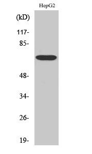
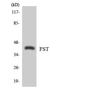
Specificity:Follistatin Polyclonal Antibody detects endogenous levels of Follistatin protein.
Formulation:Liquid in PBS containing 50% glycerol, 0.5% BSA and 0.02% sodium azide.
Source:Polyclonal, Rabbit,IgG
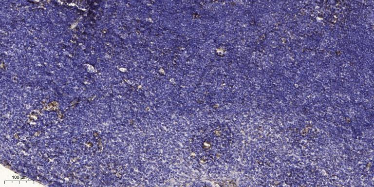
Dilution:WB 1:500 - 1:2000. IHC 1:100 - 1:300. ELISA: 1:5000.. IF 1:50-200
Purification:The antibody was affinity-purified from rabbit antiserum by affinity-chromatography using epitope-specific immunogen.
Concentration:1 mg/ml
Storage Stability:-15°C to -25°C/1 year(Do not lower than -25°C)
Other Name:FST;Follistatin;FS;Activin-binding protein
Observed Band(KD):65kD
Background: Follistatin is a single-chain gonadal protein that specifically inhibits follicle-stimulating hormone release. The single FST gene encodes two isoforms, FST317 and FST344 containing 317 and 344 amino acids respectively, resulting from alternative splicing of the precursor mRNA. In a study in which 37 candidate genes were tested for linkage and association with polycystic ovary syndrome (PCOS) or hyperandrogenemia in 150 families, evidence was found for linkage between PCOS and follistatin. [provided by RefSeq, Jul 2008],
Function:function:Binds directly to activin and functions as an activin antagonist. Specific inhibitor of the biosynthesis and secretion of pituitary follicle stimulating hormone (FSH).,similarity:Contains 1 TB (TGF-beta binding) domain.,similarity:Contains 3 follistatin-like domains.,similarity:Contains 3 Kazal-like domains.,subunit:Monomer .,tissue specificity:Isoform 1 is the predominant isoform in serum but is undetectable in follicular fluid.,
Subcellular Location:Secreted.
Expression:Isoform 1 is the predominant isoform in serum but is undetectable in follicular fluid. In the embryo, strong expression is seen in the palatal epithelia, including the medial edge epithelial and midline epithelial seam of the palatal shelves. Less pronounced expression is also seen throughout the palatal shelf and tongue mesenchyme (PubMed:31215115).
商品信息已成功复制,启研竭诚为您服务


