
Target:CHST6
Fields:Glycosaminoglycan biosynthesis - keratan sulfate
Gene Name:CHST6
Protein Name:Carbohydrate sulfotransferase 6
Human Gene Id:4166
Human Swiss Prot No:Q9GZX3
Immunogen:The antiserum was produced against synthesized peptide derived from human CHST6. AA range:331-380
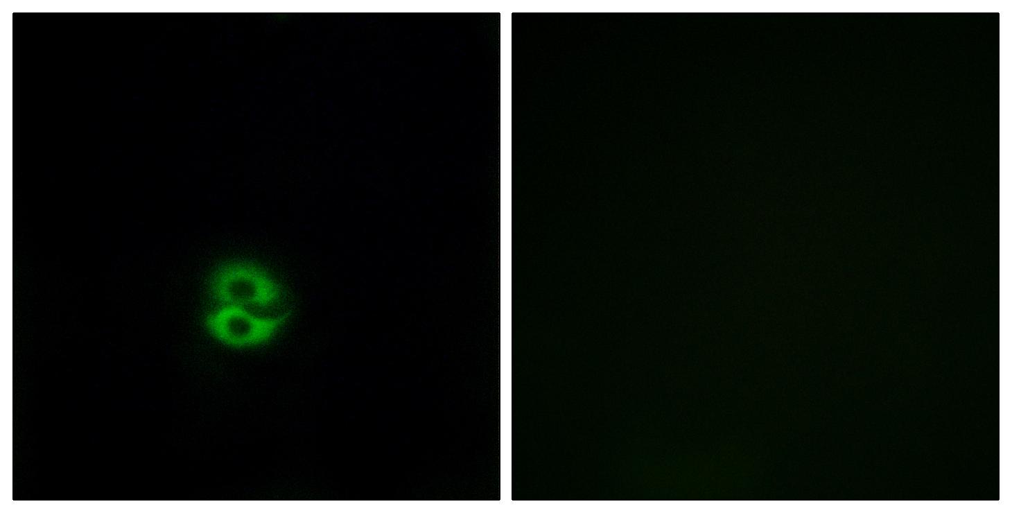
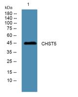
Specificity:CHST6 Polyclonal Antibody detects endogenous levels of CHST6 protein.
Formulation:Liquid in PBS containing 50% glycerol, 0.5% BSA and 0.02% sodium azide.
Source:Polyclonal, Rabbit,IgG
Dilution:IF 1:200 - 1:1000. ELISA: 1:20000. Not yet tested in other applications.
Purification:The antibody was affinity-purified from rabbit antiserum by affinity-chromatography using epitope-specific immunogen.
Concentration:1 mg/ml
Storage Stability:-15°C to -25°C/1 year(Do not lower than -25°C)
Other Name:CHST6;Carbohydrate sulfotransferase 6;Corneal N-acetylglucosamine-6-O-sulfotransferase;C-GlcNAc6ST;hCGn6ST;Galactose/N-acetylglucosamine/N-acetylglucosamine 6-O-sulfotransferase 4-beta;GST4-beta;N-acetylglucosamine 6-O-sulfotransfera
Molecular Weight(Da):44kD
Background: The protein encoded by this gene is an enzyme that catalyzes the transfer of a sulfate group to the GlcNAc residues of keratan. Keratan sulfate helps maintain corneal transparency. Defects in this gene are a cause of macular corneal dystrophy (MCD). [provided by RefSeq, Jan 2010],
Function:caution:PubMed:12824236 reported a Gly-204 variant, however according to their results reported in figure 1, it is a Gln-204 variant.,disease:Defects in CHST6 are the cause of macular corneal dystrophy (MCD) [MIM:217800]. MCD is an autosomal recessive disease characterized by corneal opacities. Onset occurs in the first decade, usually between ages 5 and 9. The disorder is progressive. Minute, gray, punctate opacities develop. Corneal sensitivity is usually reduced. Painful attacks with photophobia, foreign body sensations, and recurrent erosions occur in most patients. There are different types of MCD: MCD type I, in which there is a virtual absence of sulfated keratan sulfate (KS) in the serum and cornea, as determined by KS-specific antibodies; and MCD type II, in which the normal sulfated KS-antibody response is present in cornea and serum. MCD type I patients usually have a homozygo
Subcellular Location:Golgi apparatus membrane ; Single-pass type II membrane protein .
Expression:Expressed in cornea. Mainly expressed in brain. Also expressed in spinal cord and trachea.
商品信息已成功复制,启研竭诚为您服务


