Target:N-Shc
Fields:EGFR tyrosine kinase inhibitor resistance;Endocrine resistance;ErbB signaling pathway;Ras signaling pathway;Chemokine signaling pathway;Phospholipase D signaling pathway;Focal adhesion;Natural killer cell mediated cytotoxicity;Neurotrophin signaling pathway;Insulin signaling pathway;Estrogen signaling pathway;Prolactin signaling pathway;Relaxin signaling pathway;Growth hormone synthesis, secretion and action;Alcoholism;Bacterial invasion of epithelial cells;Glioma;Chronic myeloid leukemia;Breast cancer;Hepatocellular carcinoma;Gastric cancer
Gene Name:SHC3
Protein Name:SHC-transforming protein 3
Human Gene Id:53358
Human Swiss Prot No:Q92529
Mouse Gene Id:20418
Mouse Swiss Prot No:Q61120
Rat Swiss Prot No:O70143
Immunogen:The antiserum was produced against synthesized peptide derived from human SHC3. AA range:291-340
Specificity:N-Shc Polyclonal Antibody detects endogenous levels of N-Shc protein.
Formulation:Liquid in PBS containing 50% glycerol, 0.5% BSA and 0.02% sodium azide.
Source:Polyclonal, Rabbit,IgG
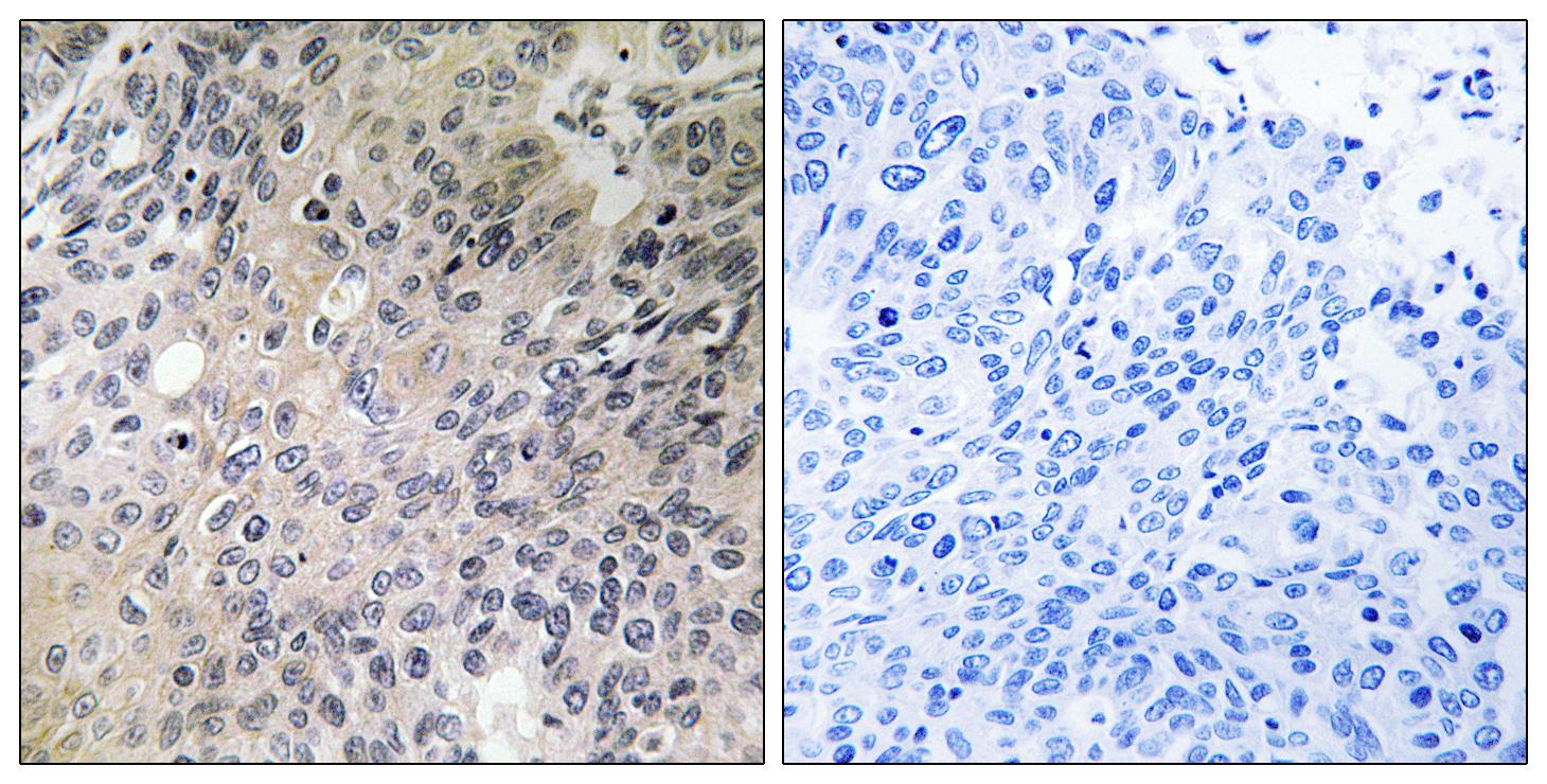
Dilution:IHC 1:100 - 1:300. ELISA: 1:10000.. IF 1:50-200
Purification:The antibody was affinity-purified from rabbit antiserum by affinity-chromatography using epitope-specific immunogen.
Concentration:1 mg/ml
Storage Stability:-15°C to -25°C/1 year(Do not lower than -25°C)
Other Name:SHC3;NSHC;SHCC;SHC-transforming protein 3;Neuronal Shc;N-Shc;Protein Rai;SHC-transforming protein C;Src homology 2 domain-containing-transforming protein C3;SH2 domain protein C3
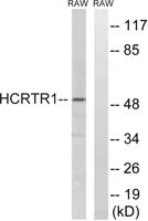
Observed Band(KD):48kD
Background:function:Signaling adapter that couples activated growth factor receptors to signaling pathway in neurons. Involved in the signal transduction pathways of neurotrophin-activated Trk receptors in cortical neurons.,PTM:Tyrosine phosphorylated.,similarity:Contains 1 PID domain.,similarity:Contains 1 SH2 domain.,subunit:Interacts with the Trk receptors in a phosphotyrosine-dependent manner. Once activated, binds to GRB2. Interacts with activated EGF receptors.,tissue specificity:Mainly expressed in brain. Hardly detectable in other tissues, except in pancreas. Highly expressed in the cerebral cortex, frontal and temporal lobes, occipital pole, hippocampus, caudate nucleus and amygdala. Expressed at low level in the cerebellum, medulla and spinal cord.,
Function:function:Signaling adapter that couples activated growth factor receptors to signaling pathway in neurons. Involved in the signal transduction pathways of neurotrophin-activated Trk receptors in cortical neurons.,PTM:Tyrosine phosphorylated.,similarity:Contains 1 PID domain.,similarity:Contains 1 SH2 domain.,subunit:Interacts with the Trk receptors in a phosphotyrosine-dependent manner. Once activated, binds to GRB2. Interacts with activated EGF receptors.,tissue specificity:Mainly expressed in brain. Hardly detectable in other tissues, except in pancreas. Highly expressed in the cerebral cortex, frontal and temporal lobes, occipital pole, hippocampus, caudate nucleus and amygdala. Expressed at low level in the cerebellum, medulla and spinal cord.,
Subcellular Location:cytosol,plasma membrane,
Expression:Mainly expressed in brain. Hardly detectable in other tissues, except in pancreas. Highly expressed in the cerebral cortex, frontal and temporal lobes, occipital pole, hippocampus, caudate nucleus and amygdala. Expressed at low level in the cerebellum, medulla and spinal cord.
商品信息已成功复制,启研竭诚为您服务