
Target:TRAC-1
Fields:RIG-I-like receptor signaling pathway
Gene Name:RNF125
Protein Name:E3 ubiquitin-protein ligase RNF125
Human Gene Id:54941
Human Swiss Prot No:Q96EQ8
Mouse Gene Id:67664
Mouse Swiss Prot No:Q9D9R0
Immunogen:The antiserum was produced against synthesized peptide derived from human RNF125. AA range:131-180
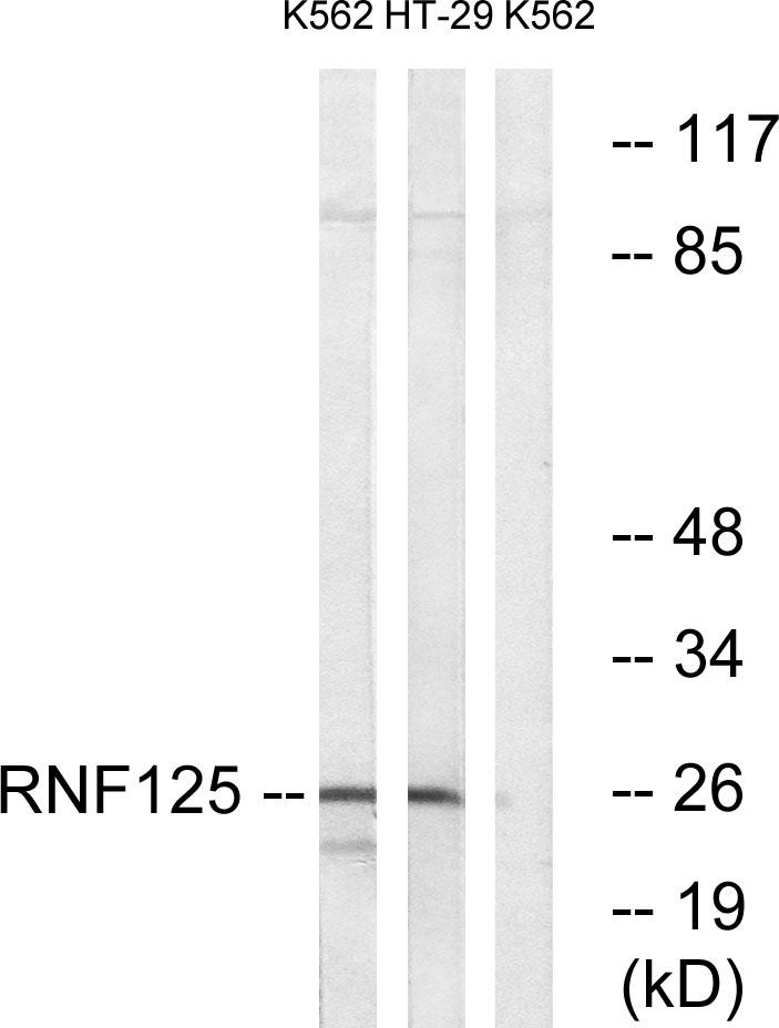
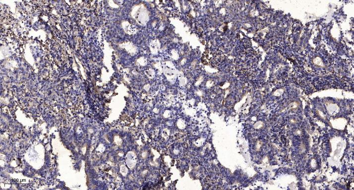
Specificity:TRAC-1 Polyclonal Antibody detects endogenous levels of TRAC-1 protein.
Formulation:Liquid in PBS containing 50% glycerol, 0.5% BSA and 0.02% sodium azide.
Source:Polyclonal, Rabbit,IgG
Dilution:WB 1:500 - 1:2000. IHC 1:100 - 1:300. ELISA: 1:10000.. IF 1:50-200
Purification:The antibody was affinity-purified from rabbit antiserum by affinity-chromatography using epitope-specific immunogen.
Concentration:1 mg/ml
Storage Stability:-15°C to -25°C/1 year(Do not lower than -25°C)
Other Name:RNF125;E3 ubiquitin-protein ligase RNF125;RING finger protein 125;T-cell RING activation protein 1;TRAC-1
Observed Band(KD):26kD
Background:ring finger protein 125(RNF125) Homo sapiens This gene encodes a novel E3 ubiquitin ligase that contains a RING finger domain in the N-terminus and three zinc-binding and one ubiquitin-interacting motif in the C-terminus. As a result of myristoylation, this protein associates with membranes and is primarily localized to intracellular membrane systems. The encoded protein may function as a positive regulator in the T-cell receptor signaling pathway. [provided by RefSeq, Mar 2012],
Function:function:E3 ubiquitin-protein ligase that acts as a positive regulator of T-cell activation. E3 ligase proteins mediate ubiquitination and subsequent proteasomal degradation of target proteins.,pathway:Protein modification; protein ubiquitination.,similarity:Contains 1 RING-type zinc finger.,tissue specificity:Predominantly expressed in lymphoid tissues, including bone marrow, spleen and thymus. Also weakly expressed in other tissues. Predominant in the CD4+ and CD8+ T-cells, suggesting that it is preferentially confined to T-cells.,
Subcellular Location:Golgi apparatus membrane ; Lipid-anchor . Shows a reticular staining pattern within the cell and is probably expressed at other intracellular membranes in addition to the Golgi membrane. Not detected at the plasma membrane. .
Expression:Predominantly expressed in lymphoid tissues, including bone marrow, spleen and thymus. Also weakly expressed in other tissues. Predominant in the CD4(+) and CD8(+) T-cells, suggesting that it is preferentially confined to T-cells.
商品信息已成功复制,启研竭诚为您服务


