
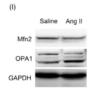
Target:OPA1
Fields:Spinocerebellar ataxia
Gene Name:OPA1 KIAA0567
Protein Name:Dynamin-like 120 kDa protein, mitochondrial (Optic atrophy protein 1) [Cleaved into: Dynamin-like 120 kDa protein, form S1]
Human Gene Id:4976
Human Swiss Prot No:O60313
Mouse Swiss Prot No:P58281
Rat Swiss Prot No:Q2TA68
Immunogen:Synthesized peptide derived from part region of human protein
Specificity:OPA1 Polyclonal Antibody detects endogenous levels of protein.
Formulation:Liquid in PBS containing 50% glycerol, and 0.02% sodium azide.
Source:Polyclonal, Rabbit,IgG
Dilution:WB 1:500-2000 ELISA 1:5000-20000
Purification:The antibody was affinity-purified from rabbit antiserum by affinity-chromatography using epitope-specific immunogen.
Concentration:1 mg/ml
Storage Stability:-15°C to -25°C/1 year(Do not lower than -25°C)
Observed Band(KD):105kD
Background: This gene product is a nuclear-encoded mitochondrial protein with similarity to dynamin-related GTPases. It is a component of the mitochondrial network. Mutations in this gene have been associated with optic atrophy type 1, which is a dominantly inherited optic neuropathy resulting in progressive loss of visual acuity, leading in many cases to legal blindness. Multiple transcript variants encoding different isoforms have been found for this gene. [provided by RefSeq, Mar 2009],
Function:disease:Defects in OPA1 are a cause of optic atrophy type 1 (OPA1) [MIM:165500]. OPA1 is a dominantly inherited optic neuropathy occurring in 1 in 50,000 individuals that features progressive loss in visual acuity leading, in many cases, to legal blindness.,disease:Defects in OPA1 are the cause of optic atrophy 1 and deafness [MIM:125250]. Some individuals with mutations in OPA1 manifest also ophthalmoplegia and myopathy.,function:Dynamin-related GTPase required for mitochondrial fusion and regulation of apoptosis. May form a diffusion barrier for proteins stored in mitochondrial cristae. Proteolytic processing in response to intrinsic apoptotic signals may lead to disassembly of OPA1 oligomers and release of the caspase activator cytochrome C (CYCS) into the mitochondrial intermembrane space.,PTM:PARL-dependent proteolytic processing releases an antiapoptotic soluble form not required f
Subcellular Location:Mitochondrion inner membrane ; Single-pass membrane protein . Mitochondrion intermembrane space . Mitochondrion membrane . Detected at contact sites between endoplasmic reticulum and mitochondrion membranes. .
Expression:Highly expressed in retina. Also expressed in brain, testis, heart and skeletal muscle. Isoform 1 expressed in retina, skeletal muscle, heart, lung, ovary, colon, thyroid gland, leukocytes and fetal brain. Isoform 2 expressed in colon, liver, kidney, thyroid gland and leukocytes. Low levels of all isoforms expressed in a variety of tissues.
商品信息已成功复制,启研竭诚为您服务


