
Target:COL8A2
Fields:Protein digestion and absorption
Gene Name:COL8A2
Protein Name:COL8A2
Human Gene Id:1296
Human Swiss Prot No:P25067
Mouse Gene Id:329941
Mouse Swiss Prot No:P25318
Immunogen:Synthesized peptide derived from human COL8A2. at AA range: 611-660
Specificity:COL8A2 Polyclonal Antibody detects endogenous levels of COL8A2
Formulation:Liquid in PBS containing 50% glycerol, 0.5% BSA and 0.02% sodium azide.
Source:Polyclonal, Rabbit,IgG
Dilution:WB 1:500-2000, ELISA 1:10000-20000
Purification:The antibody was affinity-purified from rabbit antiserum by affinity-chromatography using epitope-specific immunogen.
Concentration:1 mg/ml
Storage Stability:-15°C to -25°C/1 year(Do not lower than -25°C)
Other Name:Collagen alpha-2(VIII) chain (Endothelial collagen)
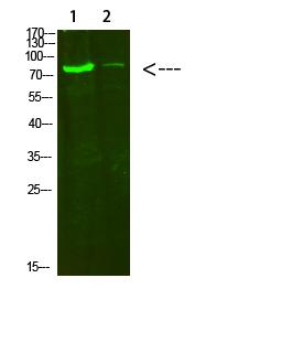
Observed Band(KD):80kD
Background: This gene encodes the alpha 2 chain of type VIII collagen. This protein is a major component of the basement membrane of the corneal endothelium and forms homo- or heterotrimers with alpha 1 (VIII) type collagens. Defects in this gene are associated with Fuchs endothelial corneal dystrophy and posterior polymorphous corneal dystrophy type 2. Alternative splicing results in multiple transcript variants. [provided by RefSeq, Jun 2014],
Function:disease:Defects in COL8A2 are a cause of Fuchs endothelial corneal dystrophy (FECD) [MIM:136800]. FECD is the commonest primary disorder of the corneal endothelium in developed countries. Symptoms of painful visual loss result from corneal decompensation. Signs may be present from the fourth decade of life onwards. Tipically, focal wart-like guttata arising from Descemet membrane develops in the central cornea; Descemet membrane is thickened by abnormal collagenous deposition. FECD is usually sporadic but familial highly penetrant forms showing autosomal dominant inheritance are also recognized.,disease:Defects in COL8A2 are a cause of posterior polymorphous corneal dystrophy (PPCD) [MIM:122000]. PPCD is a slowly progressive hereditary disorder of the corneal endothelium that leads to a variable degree of visual impairment usually in adulthood. PPCD is usually inherited as an autosomal d
Subcellular Location:Secreted, extracellular space, extracellular matrix, basement membrane.
Expression:Expressed primarily in the subendothelium of large blood vessels. Also expressed in arterioles and venules in muscle, heart, kidney, spleen, umbilical cord, liver and lung and is also found in connective tissue layers around hair follicles, around nerve bundles in muscle, in the dura of the optic nerve, in cornea and sclera, and in the perichondrium of cartilaginous tissues. In the kidney, expressed in mesangial cells, glomerular endothelial cells, and tubular epithelial cells. Also expressed in mast cells, and in astrocytes during the repair process. Expressed in Descemet's membrane.
商品信息已成功复制,启研竭诚为您服务


