
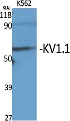
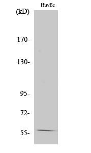
Target:KV1.1
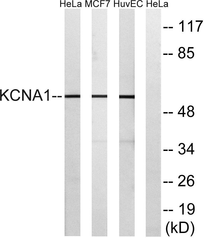
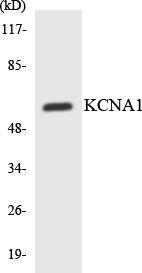
Gene Name:KCNA1
Protein Name:Potassium voltage-gated channel subfamily A member 1
Human Gene Id:3736
Human Swiss Prot No:Q09470
Mouse Gene Id:16485
Mouse Swiss Prot No:P16388
Rat Gene Id:24520
Rat Swiss Prot No:P10499
Immunogen:The antiserum was produced against synthesized peptide derived from human KCNA1. AA range:256-305
Specificity:KV1.1 Polyclonal Antibody detects endogenous levels of KV1.1 protein.
Formulation:Liquid in PBS containing 50% glycerol, 0.5% BSA and 0.02% sodium azide.
Source:Polyclonal, Rabbit,IgG
Dilution:WB 1:500-2000;IHC 1:50-300; ELISA 2000-20000
Purification:The antibody was affinity-purified from rabbit antiserum by affinity-chromatography using epitope-specific immunogen.
Concentration:1 mg/ml
Storage Stability:-15°C to -25°C/1 year(Do not lower than -25°C)
Other Name:KCNA1;Potassium voltage-gated channel subfamily A member 1;Voltage-gated K(+) channel HuKI;Voltage-gated potassium channel HBK1;Voltage-gated potassium channel subunit Kv1.1
Observed Band(KD):57kD
Background: This gene encodes a voltage-gated delayed potassium channel that is phylogenetically related to the Drosophila Shaker channel. The encoded protein has six putative transmembrane segments (S1-S6), and the loop between S5 and S6 forms the pore and contains the conserved selectivity filter motif (GYGD). The functional channel is a homotetramer. The N-terminus of the channel is associated with beta subunits that can modify the inactivation properties of the channel as well as affect expression levels. The C-terminus of the channel is complexed to a PDZ domain protein that is responsible for channel targeting. Mutations in this gene have been associated with myokymia with periodic ataxia (AEMK). [provided by RefSeq, Jul 2008],
Function:disease:Defects in KCNA1 are the cause of episodic ataxia type 1 (EA1) [MIM:160120]; also known as paroxysmal or episodic ataxia with myokymia (EAM) or paroxysmal ataxia with neuromyotonia. EA1 is an autosomal dominant disorder characterized by brief episodes of ataxia and dysarthria. Neurological examination during and between the attacks demonstrates spontaneous, repetitive discharges in the distal musculature (myokymia) that arise from peripheral nerve. Nystagmus is absent.,disease:Defects in KCNA1 are the cause of myokymia isolated type 1 (MK1) [MIM:160120]. Myokymia is a condition characterized by spontaneous involuntary contraction of muscle fiber groups that can be observed as vermiform movement of the overlying skin. Electromyography typically shows continuous motor unit activity with spontaneous oligo- and multiplet-discharges of high intraburst frequency (myokymic discharges).
Subcellular Location:Cell membrane ; Multi-pass membrane protein . Membrane . Cell projection, axon . Cytoplasmic vesicle . Perikaryon . Endoplasmic reticulum . Cell projection, dendrite . Cell junction . Cell junction, synapse . Cell junction, synapse, presynaptic cell membrane . Cell junction, synapse, presynapse . Homotetrameric KCNA1 is primarily located in the endoplasmic reticulum. Interaction with KCNA2 and KCNAB2 or with KCNA4 and KCNAB2 promotes expression at the cell membrane (By similarity). .
Expression:Detected adjacent to nodes of Ranvier in juxtaparanodal zones in spinal cord nerve fibers, but also in paranodal regions in some myelinated spinal cord axons (at protein level) (PubMed:11086297). Detected in the islet of Langerhans (PubMed:21483673).
商品信息已成功复制,启研竭诚为您服务


