
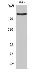
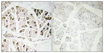
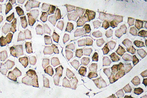
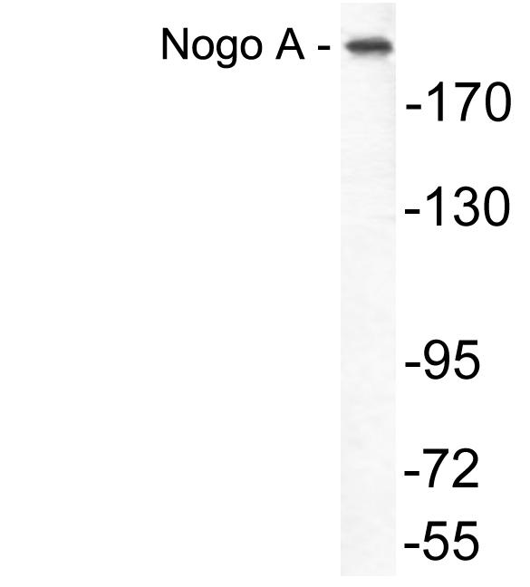
Target:Nogo A
Fields:Alzheimer disease
Gene Name:RTN4
Protein Name:Reticulon-4
Human Gene Id:57142
Human Swiss Prot No:Q9NQC3
Mouse Gene Id:68585
Mouse Swiss Prot No:Q99P72
Rat Gene Id:83765
Rat Swiss Prot No:Q9JK11
Immunogen:The antiserum was produced against synthesized peptide derived from human Nogo A. AA range:450-499
Specificity:Nogo A Polyclonal Antibody detects endogenous levels of Nogo A protein.
Formulation:Liquid in PBS containing 50% glycerol, 0.5% BSA and 0.02% sodium azide.
Source:Polyclonal, Rabbit,IgG
Dilution:WB 1:500 - 1:2000. IHC 1:100 - 1:300. ELISA: 1:10000.
Purification:The antibody was affinity-purified from rabbit antiserum by affinity-chromatography using epitope-specific immunogen.
Concentration:1 mg/ml
Storage Stability:-15°C to -25°C/1 year(Do not lower than -25°C)
Other Name:RTN4;KIAA0886;NOGO;My043;SP1507;Reticulon-4;Foocen;Neurite outgrowth inhibitor;Nogo protein;Neuroendocrine-specific protein;NSP;Neuroendocrine-specific protein C homolog;RTN-x;Reticulon-5
Observed Band(KD):220kD
Background: This gene belongs to the family of reticulon encoding genes. Reticulons are associated with the endoplasmic reticulum, and are involved in neuroendocrine secretion or in membrane trafficking in neuroendocrine cells. The product of this gene is a potent neurite outgrowth inhibitor which may also help block the regeneration of the central nervous system in higher vertebrates. Alternatively spliced transcript variants derived both from differential splicing and differential promoter usage and encoding different isoforms have been identified. [provided by RefSeq, Jul 2008],
Function:domain:Three regions, residues 59-172, 544-725 and the loop 66 amino acids, between the two transmembrane domains, known as Nogo-66 loop, appear to be responsible for the inhibitory effect on neurite outgrowth and the spreading of neurons. This Nogo-66 loop, mediates also the binding of RTN4 to its receptor.,function:Potent neurite growth inhibitor in vitro and plays a role both in the restriction of axonal regeneration after injury and in structural plasticity in the CNS. Isoform 2 reduces the anti-apoptotic activity of Bcl-xl and Bcl-2. This is likely consecutive to their change in subcellular location, from the mitochondria to the endoplasmic reticulum, after binding and sequestration. Isoform 2 and isoform 3 inhibit BACE1 activity and amyloid precursor protein processing.,online information:Nerve regrowth: nipped by a no-go - Issue 69 of April 2006,online information:The Singapore hu
Subcellular Location:[Isoform A]: Endoplasmic reticulum membrane ; Multi-pass membrane protein . Cell membrane; Multi-pass membrane protein ; Cytoplasmic side . Anchored to the membrane of the endoplasmic reticulum (ER) through 2 putative transmembrane domains. Localizes throughout the ER tubular network (PubMed:27619977). Co-localizes with TMEM33 at the ER sheets. .; [Isoform B]: Endoplasmic reticulum membrane ; Multi-pass membrane protein . Cell membrane ; Multi-pass membrane protein ; Extracellular side . Cell junction . Mainly located on endoplasmic reticulum tubules and sheet edges (PubMed:27786289). Upon ICAM1 engagement, redistributed toward endothelial junctions where interacts with CDH5 (PubMed:21183689). .; [Isoform C]: Endoplasmic reticulum membrane ; Multi-pass membrane protein .
Expression:Isoform A: is specifically expressed in brain and testis and weakly in heart and skeletal muscle. Isoform B: widely expressed except for the liver. Highly expressed in endothelial cells and vascular smooth muscle cells, including blood vessels and mesenteric arteries (PubMed:15034570, PubMed:21183689). Isoform C: is expressed in brain, skeletal muscle and adipocytes. Isoform D is testis-specific.
商品信息已成功复制,启研竭诚为您服务


